24/7 Customer Assistance
24/7 Assistance with the best medical care at your service

Call us for any question
+91 90858 83067BLOCK-B, PH-1, Sushant Lok Phase I, Gurugram, Haryana 122001
IndiaOffice Hour: 09:00am - 09:00pm
info@medigocare.com
Published: June 2026
Diabetic retinopathy is damage to the retinal blood vessels caused by long-term high blood sugar. It is a leading cause of preventable vision loss — and in its early stages it causes no symptoms at all, which is why a yearly retinal examination is essential for every person with diabetes, regardless of how well their vision feels.
Diabetes is a disease that most people understand in terms of blood sugar, insulin and diet. Fewer realise that the damage it does to blood vessels spreads far beyond the pancreas — and that the eyes are among the most vulnerable organs it silently affects. Diabetic retinopathy, the name for this eye damage, develops in a majority of people who live with diabetes long enough. It progresses without pain. It produces no symptoms in the early stages. And by the time vision changes are obvious, the window for the easiest treatment may already have closed.
The scale of this problem is significant. According to the SMART-India national screening study published in The Lancet Global Health, based on the estimated 101 million people with diabetes in India in 2021, approximately 21 million people with diabetes have vision impairment, of whom 2.4 million are blind. Globally, the International Diabetes Federation projects that the number of people with diabetes will rise from 537 million in 2021 to 643 million by 2030 and 783 million by 2045 — and the burden of diabetic retinopathy will grow in parallel, particularly in low- and middle-income countries where screening is least structured.
Understanding how diabetic retinopathy develops, what its warning signs look like and when those signs mean you need same-day medical attention is not a specialist concern. It is essential knowledge for anyone with diabetes, and for the families and healthcare providers who care for them.
The retina is the thin layer of light-sensitive tissue at the back of your eye. It is one of the most metabolically demanding tissues in the body, supplied by a dense network of tiny blood vessels. Chronically elevated blood sugar — the defining feature of poorly controlled diabetes — damages those vessels in two specific ways.
First, it makes the vessel walls fragile and leaky. Fluid and blood seep out of weakened vessels into the surrounding retinal tissue, disrupting the precise optical environment the retina needs to function. When this leakage reaches the macula — the central, high-resolution zone of the retina — vision blurs.
Second, some damaged vessels eventually block completely. When patches of retina lose their blood supply, the body responds by growing new vessels to compensate. But these new vessels are structurally abnormal: fragile, disorganised and prone to bleeding. Their growth marks the transition to the advanced stage of diabetic retinopathy — and their consequences, including vitreous haemorrhage and retinal detachment, can cause rapid, severe vision loss.
The insidious aspect of this process is its silence. Retinal vessel damage can be extensive before any vision change occurs. According to the SMART-India study, 2.4% of people newly diagnosed with diabetes in India already have vision-threatening retinopathy at the time of diagnosis — meaning the damage had been accumulating undetected while the diabetes itself went undiagnosed. That single statistic makes the case for retinal screening at the time of diabetes diagnosis, not only in established diabetes.
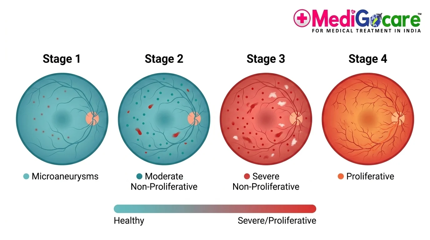 Diagram: Four stages of diabetic retinopathy from mild microaneurysms to proliferative new vessels.
The honest starting point: diabetic retinopathy often has no symptoms in its early stages. Damage is occurring at a microscopic level in the vessel walls and retinal tissue that you cannot feel and that does not affect what you see — until enough damage has accumulated to affect vision. This is precisely why screening matters. An ophthalmologist examining a dilated retina or reviewing a retinal photograph can detect the earliest changes — microaneurysms, haemorrhages, leaking vessels — long before any symptom appears.
During the first two stages of diabetic retinopathy (mild and moderate non-proliferative disease), most people experience no changes in their sight whatsoever. Vision may feel completely normal. This is the window when the condition is most treatable — but since the patient feels nothing, it is also the window most commonly missed. A yearly dilated retinal examination or retinal photograph is the only reliable way to catch it here.
The single most important action any person with diabetes can take for their eye health is attending a dilated retinal examination at least once a year, starting from the time of diagnosis. Not when vision blurs. Not when dark spots appear. Every year.
When symptoms do appear, they fall into several recognisable patterns. Understanding what each one means helps you decide how urgently to act.
Blurry or fluctuating vision comes in two distinct forms. Short-term blur — vision that is hazy one day and clearer the next, often correlating with blood sugar swings — happens because changing glucose levels alter the shape of the eye's natural lens. This is annoying but not dangerous on its own. More concerning is persistent central blur, which typically signals diabetic macular edema (see below): fluid accumulating beneath the macula, causing the central high-resolution zone to swell and distort. This kind of blur doesn't fluctuate with sugar levels — it is more consistent, and it requires assessment.
Floaters and dark spots — dark strings, dots or cobweb shapes that drift across your vision — are one of the more alarming symptoms because of what they may represent. Small numbers of floaters are common and often benign in the general population. In a person with diabetes, particularly one with poor long-term blood sugar control, new or increased floaters are more likely to signal bleeding from fragile retinal vessels into the vitreous gel. This is a sign of advanced, proliferative diabetic retinopathy, and it warrants prompt specialist assessment — not watchful waiting.
Patches of missing or darkened vision — gaps, dark areas or a shadow in a specific part of the visual field — can indicate areas of retinal ischaemia (patches of retina that have lost their blood supply) or more serious bleeding.
Poor night vision and reduced colour contrast — early deterioration in low-light performance, or colours that appear washed out or less vivid than they used to — can reflect diffuse retinal damage before obvious structural changes appear on examination.
 Simulation: Visual symptoms indicating blurry vision, floaters, and dark spots.
Some symptoms are not "monitor and review" situations. They require same-day assessment by an ophthalmologist:
If you experience any of these, do not wait for a routine appointment. Contact an eye emergency service or attend an emergency ophthalmology clinic immediately. Retinal detachment left untreated for even a few hours can result in permanent blindness that cannot be reversed.
The severity of diabetic retinopathy is classified using the Early Treatment Diabetic Retinopathy Study (ETDRS) system, which categorises the condition into five levels — no DR, mild NPDR, moderate NPDR, severe NPDR, and PDR. For practical clinical use, four stages are commonly described to patients:
Stage 1 — Mild Non-Proliferative Diabetic Retinopathy (NPDR)
A small number of microaneurysms — tiny balloon-like bulges in retinal blood vessel walls — are visible on fundus examination. These are the earliest detectable sign of diabetic retinal damage. Vision is typically completely normal. The retina is beginning to show the effects of prolonged high blood sugar, but structural function is preserved.
What to do: attend the ophthalmology review your doctor recommends (annually for most patients at this stage), optimise blood glucose, blood pressure and blood fats, and do not miss follow-up appointments. The evidence from the ETDRS is clear — good glycaemic control at this stage genuinely slows progression.
Stage 2 — Moderate NPDR
More widespread vessel damage: more numerous microaneurysms, intraretinal haemorrhages (small bleeds within the retinal layers), hard exudates (yellowish deposits of leaked lipids) and possible areas of retinal thickening. Vision may still be normal or near-normal, but if macular edema has developed, some blurring of central vision may be noticed. Review intervals shorten — typically six-monthly.
What to do: keep all ophthalmology appointments. If macular edema is present, treatment with anti-VEGF injections begins at this stage. Do not delay treatment if it is recommended.
Stage 3 — Severe NPDR
The retina is under significant stress. Extensive intraretinal haemorrhages appear in all four quadrants of the retina, venous beading (irregular, varicose-like swelling of retinal veins) is present, and there is significant intraretinal microvascular abnormality. The risk of progressing to proliferative disease within the next year is approximately 15% in one eye and 25% in both eyes. Vision may still be functional, but this stage requires close, frequent monitoring.
What to do: ophthalmology reviews every three to four months. Pan-retinal photocoagulation (laser) may be considered to reduce the risk of progression. If macular edema is worsening, anti-VEGF treatment intensifies.
Stage 4 — Proliferative Diabetic Retinopathy (PDR)
New, abnormal blood vessels (neovascularisation) have grown on the surface of the retina or on the optic disc. These fragile new vessels can bleed into the vitreous — causing a sudden onset of floaters, significant darkening of vision or complete obscuration. They can also trigger scar tissue formation that pulls on the retina, causing tractional retinal detachment. Proliferative retinopathy is sight-threatening and requires active treatment as a priority.
What to do: do not delay. Pan-retinal laser photocoagulation and/or intravitreal anti-VEGF injections are the primary treatments for PDR. Vitrectomy surgery is required when vitreous haemorrhage or traction has already occurred. The sooner treatment is initiated in PDR, the better the outcome — surgery after prolonged haemorrhage or macular detachment carries less certain visual recovery than treatment before these complications develop.
Diabetic macular edema (DME) deserves its own section because it is distinct from the four-stage classification and can occur at any stage of diabetic retinopathy, including early NPDR. It is the single most common cause of vision loss in people with diabetes who do not have advanced proliferative disease.
The macula is the central, densest part of the retina responsible for the sharp vision you use for reading, recognising faces, and seeing fine detail. When vessels in and around the macula leak fluid — as they do in diabetic retinopathy — that fluid accumulates under and within the macula, causing it to thicken and swell. The result is a specific kind of blur: central vision becomes distorted, hazy or simply less sharp, while peripheral vision remains relatively intact.
The landmark clinical indicator for DME is the presence of retinal thickening on OCT (optical coherence tomography) — a fast, non-invasive scan that produces a cross-sectional image of the retinal layers. An OCT scan can detect macular swelling long before significant visual loss occurs, which is why it forms part of the standard diabetic retinal assessment at most specialist centres.
Treatment of DME is now well-established. Anti-VEGF injections — delivered directly into the vitreous of the eye in a brief outpatient procedure under topical anaesthetic — reduce the leakage and swelling by blocking the chemical signals that drive abnormal vessel permeability. Injections are typically given monthly for an initial loading period, then at extended intervals based on the eye's response. The RISE and RIDE clinical trials, which assessed ranibizumab (Lucentis) for DME, found that patients receiving monthly injections gained an average of ten or more letters of visual acuity over two years compared to sham-treated patients. Vision can genuinely improve with treatment, not merely stabilise — which is not the case for most other forms of retinal damage.
 Diagram: Diabetic macular edema (DME) fluid swelling the macula causing central blurred vision.
Treatment varies by stage and by whether macular edema is present. The good news: treatment works well when started at the right time.
Blood sugar and blood pressure control remain the most powerful tool available at every stage. Multiple major clinical trials — including the landmark DCCT (Diabetes Control and Complications Trial) for type 1 diabetes and the UKPDS for type 2 — have demonstrated that tight glycaemic control dramatically reduces both the incidence and progression of diabetic retinopathy. Lowering HbA1c by even one percentage point has measurable effects on retinopathy risk. Blood pressure control also has an independent protective effect on the retinal vasculature.
Anti-VEGF injections are the first-line treatment for diabetic macular edema and are also used in proliferative diabetic retinopathy. Bevacizumab (Avastin), ranibizumab (Lucentis) and aflibercept (Eylea) are the agents most commonly used. Each injection takes approximately 15 minutes in a clinic setting. Patients feel mild pressure but no pain — the eye is numbed with topical anaesthetic drops beforehand. A treatment course typically involves monthly injections for three to six months, then monitoring with injections as needed.
Laser photocoagulation is delivered in two forms. Focal or grid laser targets specific leaking vessels around the macula in certain forms of DME. Panretinal photocoagulation (PRP) applies laser burns across the peripheral retina to reduce the drive for abnormal new-vessel growth in proliferative retinopathy — one of the most effective ways to prevent vitreous haemorrhage and traction when PDR is detected before bleeding has occurred.
Vitrectomy surgery removes the vitreous gel from the centre of the eye when it has been significantly infiltrated by blood, or when scar tissue is pulling on the retina causing tractional detachment. It is the surgical intervention of last resort in DR — necessary and effective, but better avoided through earlier treatment.
For international patients considering treatment in India — where the same laser systems, anti-VEGF medications and vitreoretinal surgical technology are available at a fraction of Western costs — our guide to diabetic eye care in India covers the full treatment pathway, cost breakdown and how the logistics of travelling for retinal treatment work in practice.
The clinical case for acting early is not just theoretical. The outcome numbers make it concrete.
Research has found that people who have undergone treatment for vitreous haemorrhage experience around 90% vision improvement. For those with proliferative diabetic retinopathy who require surgical intervention at a more advanced stage, most experience 30% to 60% improvement in vision — a meaningful difference that directly reflects how much more damage has already occurred before treatment begins.
The same contrast applies to treatment complexity and cost. Early-stage macular edema responds to anti-VEGF injections — outpatient procedures done without a surgical theatre, with same-day recovery. Advanced proliferative disease with traction requires vitrectomy — a surgical procedure with a longer recovery, a longer hospital stay and significantly higher cost. The procedure that costs ₹20,000–₹60,000 per injection at early stage becomes ₹60,000–₹2,50,000 for vitrectomy when the same disease reaches a point that injections alone can no longer manage.
Recent research projections make this more urgent, not less. A Chang Eye Group analysis from April 2026 notes that vision-threatening diabetic retinopathy cases are expected to nearly triple by 2050 as global diabetes rates continue their upward trajectory. In India specifically, the shift toward earlier onset of diabetes — often diagnosed in the 30s and 40s rather than the 60s — means a longer lifetime exposure to elevated blood sugar and a longer window over which retinopathy can develop and progress.
The lesson is not frightening. It is empowering. The same condition that causes blindness when caught late causes only a need for monitoring when caught early, and responds to injections when caught at the macular edema stage. The tools to preserve vision exist. What they require is the window — and the window is kept open by annual retinal screening.
 Infographic: Treatment outcomes for early vs late intervention in diabetic retinopathy.
If you have diabetes and have not had a retinal examination in the last twelve months, that is the single most important action you can take today. Book a dilated retinal examination with an ophthalmologist — not just a standard optometry check — and ask for a retinal photograph or OCT if available. If you are in a country where specialist retinal care is difficult to access, or where the cost is prohibitive, India's retinal specialists and screening infrastructure offer a high-quality, internationally benchmarked alternative at a fraction of the cost of Western private care.
Send your most recent retinal reports — or simply tell us you need an initial retinal screening — on WhatsApp and we will arrange a specialist review, either remotely or through a visit to India with MediGoCare.
WhatsApp Us (+91 90858 83067)Often there are none — which is the critical point. In the early stages, diabetic retinopathy causes no symptoms and does not change what you see. As it progresses: blurred vision (particularly centrally), floaters or dark spots, poor night vision, faded colours, or a shadow over part of the visual field. Because early disease is silent, annual retinal screening is essential for every person with diabetes, regardless of how good their vision feels.
The damage already done to retinal blood vessels is not always reversible — scarring and vessel loss are permanent. However, the progression of diabetic retinopathy can almost always be halted, and in the case of macular edema, vision can genuinely improve with anti-VEGF treatment. Treatment protects the vision you still have; it is most effective when started early, before structural damage has become advanced.
Two mechanisms cause blur in diabetic patients. Short-term: swings in blood sugar alter the shape of the natural lens, causing temporary vision fluctuations. More seriously: diabetic macular edema — fluid leaking from damaged retinal vessels into the macula — causes persistent central blur. Persistent or worsening blur in a person with diabetes should always be assessed by an ophthalmologist promptly, not monitored at home.
Dark spots or floaters in a person with diabetes most often signal bleeding from fragile abnormal blood vessels (neovascularisation) into the vitreous gel — a sign of proliferative diabetic retinopathy. Even small numbers of new, sudden floaters in a diabetic patient warrant prompt assessment. A sudden large increase in floaters or a curtain of darkness across vision is an emergency requiring same-day care.
At least once a year, starting from the time of diabetes diagnosis — and regardless of how well vision feels. For patients with any existing retinopathy, or with poorly controlled blood sugar or blood pressure, review frequency increases to every six months or more. National and international guidelines from the AAO, NEI and WHO all recommend annual dilated retinal examination from the point of diabetes diagnosis.
The condition itself is not always an emergency — but some of its symptoms are. Sudden onset of many new floaters, flashing lights, or a curtain or shadow moving across vision may signal vitreous haemorrhage or retinal detachment. These require same-day emergency ophthalmological assessment. Do not wait for a scheduled appointment if any of these symptoms develop. Retinal detachment left untreated for hours carries a high risk of permanent vision loss that cannot be recovered.
24/7 Assistance with the best medical care at your service
Quality treatment with professional & unmatched personal care
Our experts make your Communication easier and more transparent

